Digital imaging and X-rays
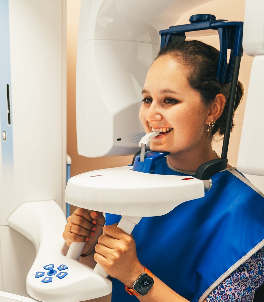
All our X-rays are digital: the image appears instantly on screen, we can enlarge it to explain it to you, and it is stored in your patient file. Depending on what we need to examine, we use three types of X-rays.
Intraoral X-ray
A small sensor placed in the mouth gives a close-up of two or three teeth. It is the basic examination to detect decay, check a root or follow up on treatment.
Panoramic X-ray
A single image of the whole mouth: teeth, jaws and joints. It provides an overview, useful at a first appointment or a check-up.
3D X-ray (CBCT)
Cone beam imaging reconstructs your jaw in three dimensions. We can navigate virtually through bone and roots, see the sinuses and nerves, and measure bone volume precisely. This examination is essential to prepare an implant placement or complex treatment.
Learn more about our expertise
-
 A fully digital practice
-
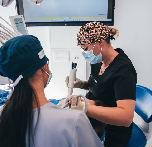 Cameras and digital impressions
-
 Commitment to quality, Dr. Frédéric DIENER, dentist in Saint-Louis
-
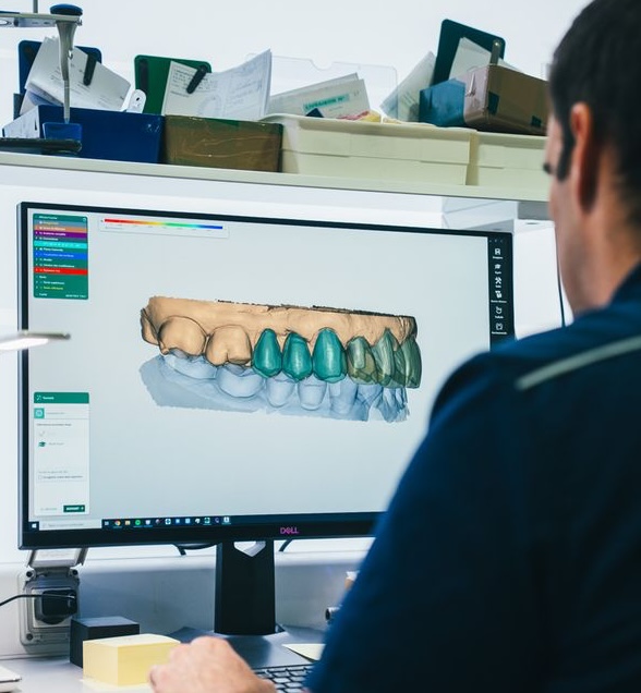 Digital design and manufacturing
-
 DR. DIENER and his team
-
 Hygiene and instrument processing
-
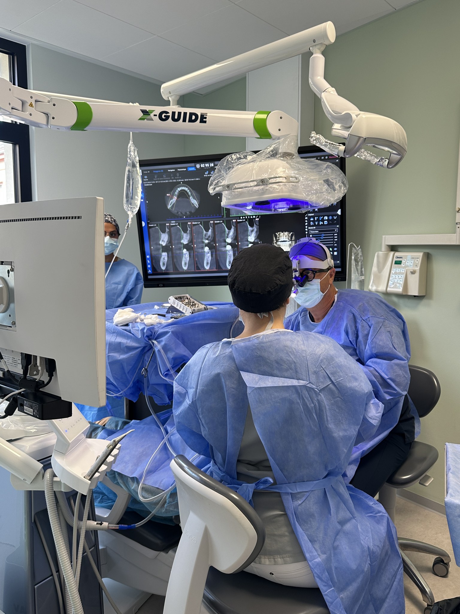 Navigated guided surgery
-
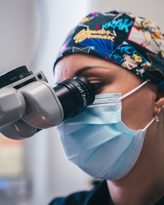 Optical aids and Zeiss microscope
-
 Patient management at Dr. Frédéric DIENER's dental practice
-
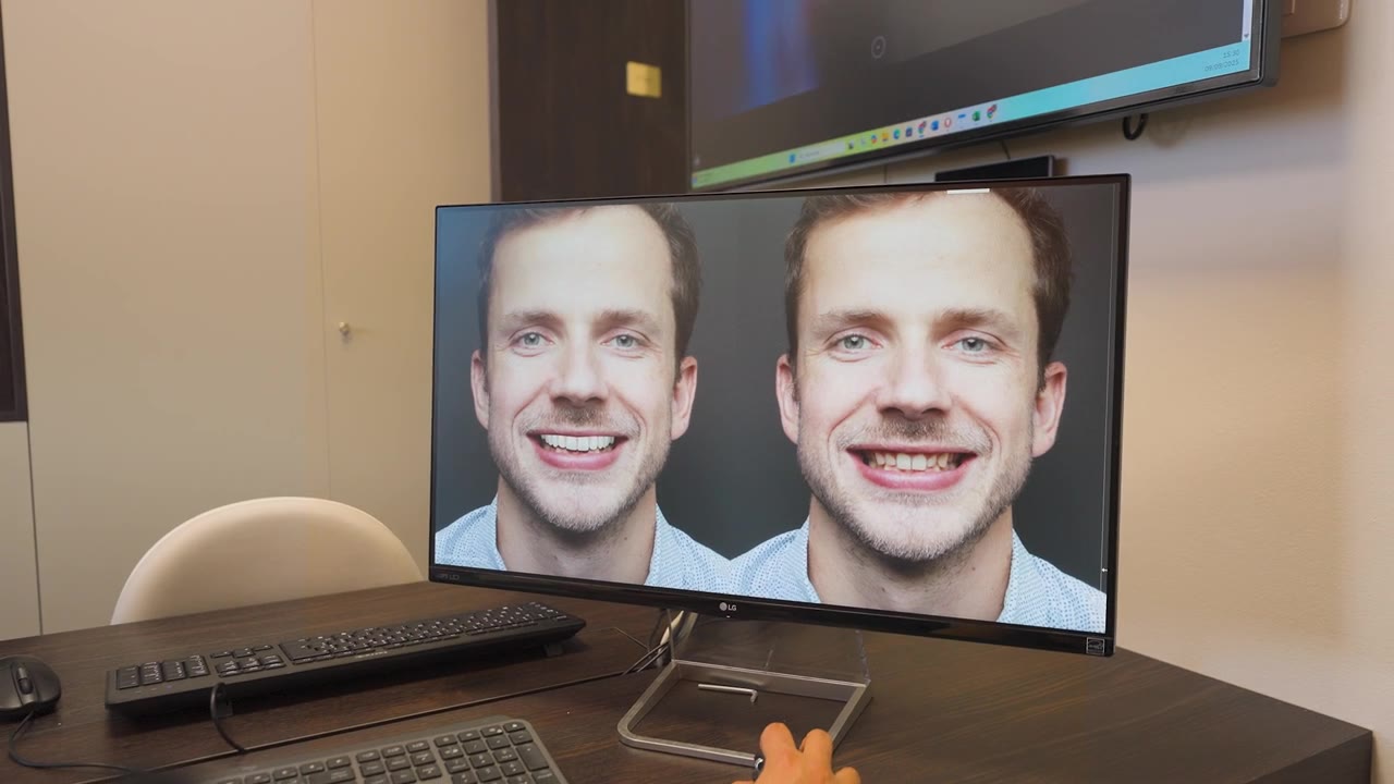 Planning and simulation
-
 The history of our dental practice